ANSTO- ROTARY CLUB OF CANTERBURY VISIT TO SYNCHROTRON
INTRODUCTION
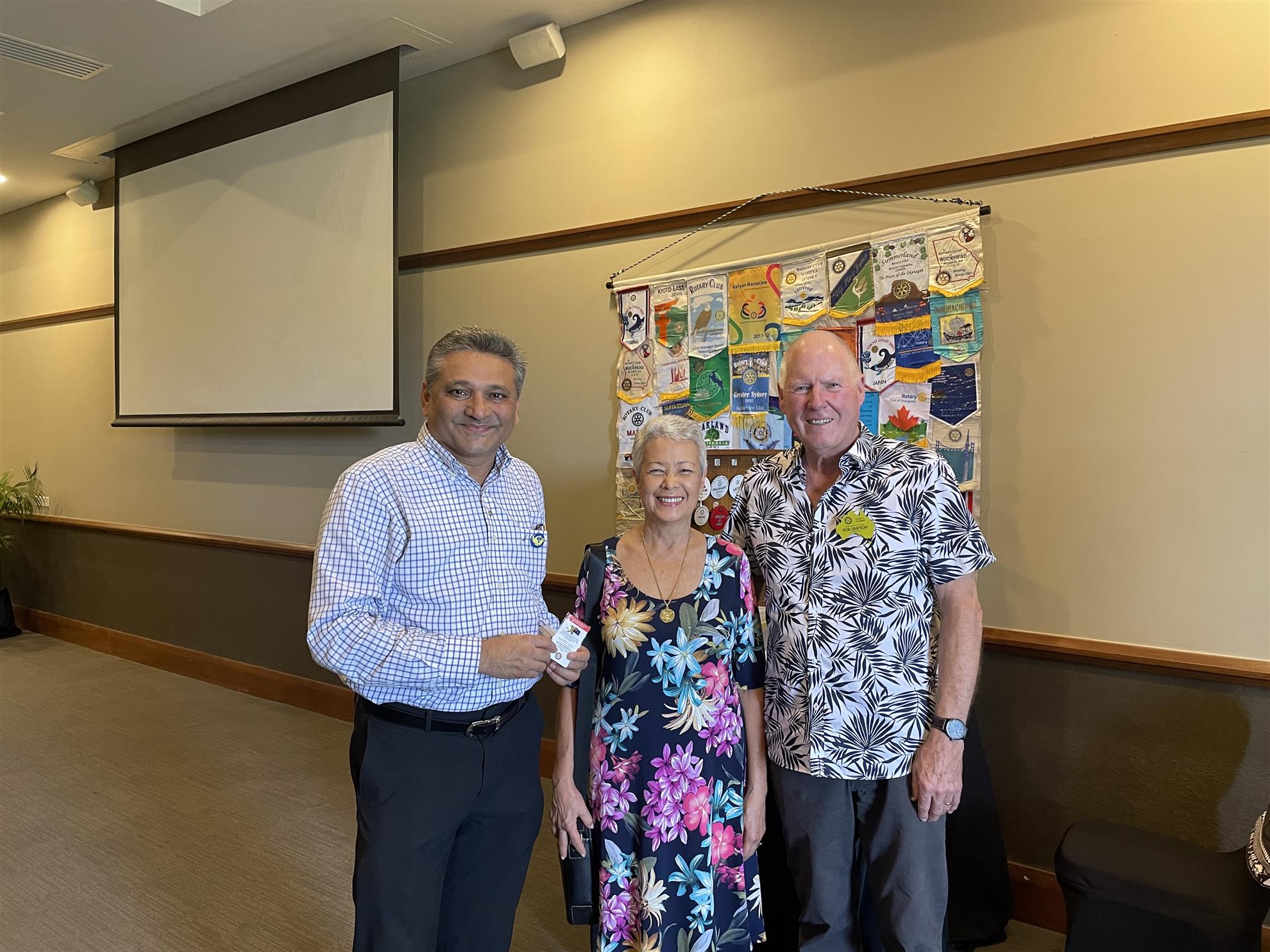
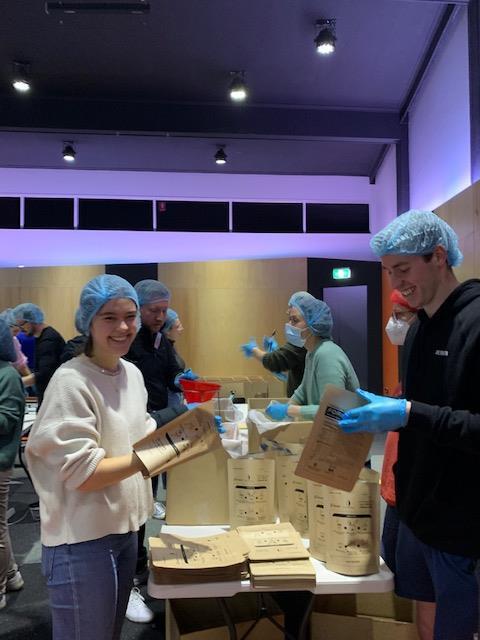
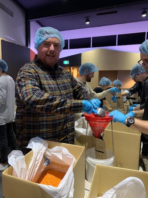
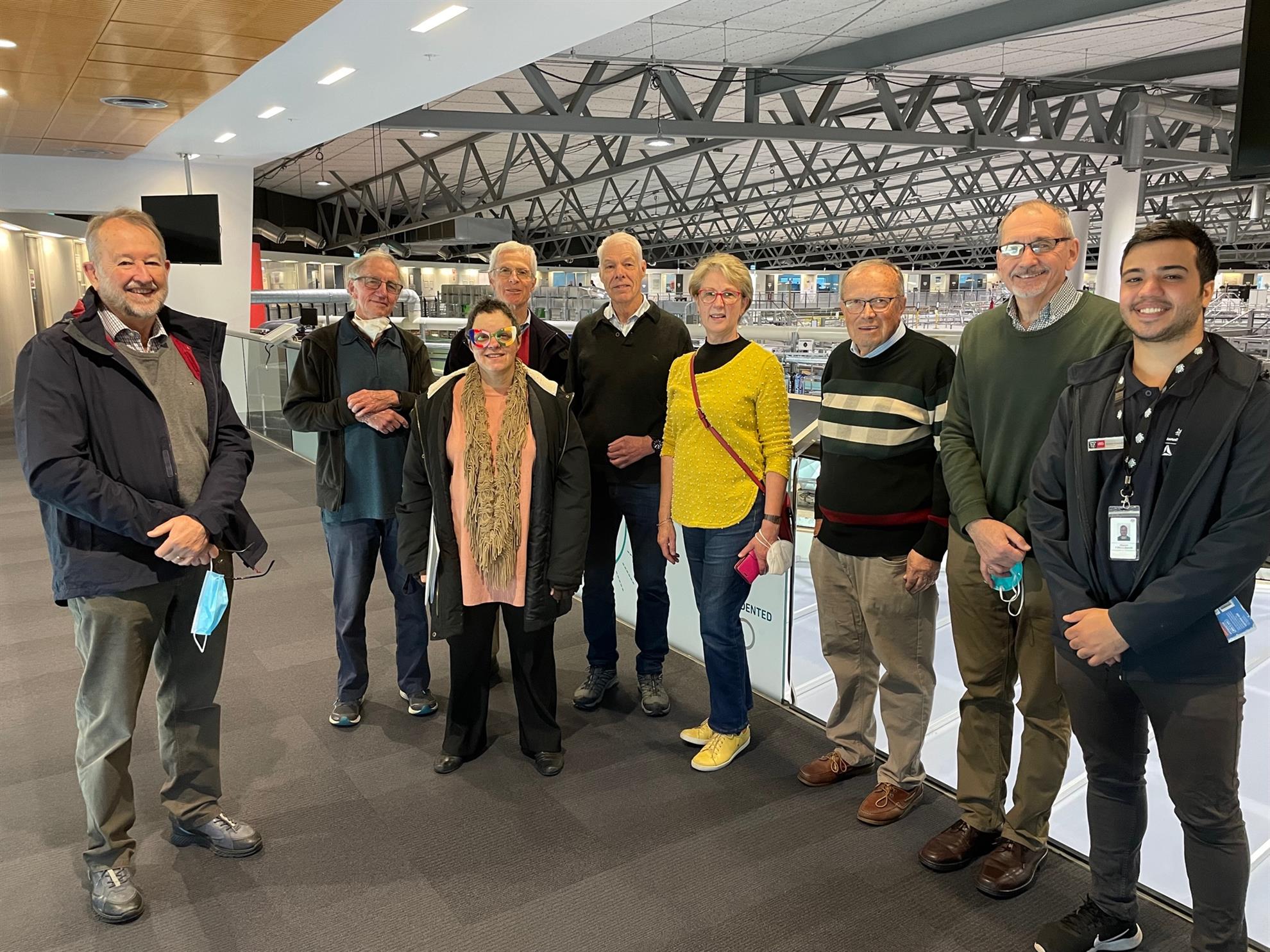
Nine members of The Rotary Club of Canterbury- Alan Stevens, Anne Josefsberg, Kyle Wightman, Neil Williams, Edda Williams, John Pocock, David Zrna, Doug Hawley and Ted Waghorne, attended our vocational visit to the Synchrotron, in Blackburn Road, Notting Hill.
Maysam, our tour guide and Education Officer, delivered a passionate and detailed tour of the Synchrotron. We began at the reception area, where he showed us the overview of the Synchrotron, using a model. Many researchers and organizations use the Synchrotron for a variety of research activities.
ENERGY
Magnets are used to create electric fields, storing energy and creating electron beam lines. A beam is a burst of energy, which is used in beam lines. Ten thousand scientists come to the location every year. These include medical researchers, geologists, mechanical engineers and a variety of other individuals. Samples are used which create images one million times brighter than the sun. The only other Synchrotron in the Southern Hemisphere is in Brazil. the images revealed as so accurate that they include soft tissue. On a map of the world, pins have been placed showing where all the researchers come from, to visit Melbourne, to use the Synchrotron. It requires lots of effort to keep the the Synchrotron running. Below the grey area of the inner workings, one inch of rubber is placed to reduce the vibrations from the nearby train station. The purpose of the location is to create a live image for research and medical purposes.
PRACTICAL OUTCOMES
Nearly one hundred percent of the images lead to changes in medical treatment in hospitals. For instance, the manner in which lung capacity of premature babies is treated. has been significantly advanced as a result of one of the live images. Paleantologists use the live images to check for specimens in rocks.
RESEARCH IS SHARED
While the Synchrotron began with four beam lines, it now has ten, and will expand to more in the future. One area, X-Ray Fluorescence Microscopy creates a signature for each atom. The example of agricultural scientists testing seeds, to highlight the metals to be able to label a food as "super" food, is one of he applications of the live image.
MAYSAM'S AREA OF INTEREST
Maysam works in the medical area of cancer therapy. He told us about scientifically analysing bush medicine, using the live images to see the exact structure of the protein, which would then provide compounds for use in cancer treatment in the body. The native habitat produces different compounds.
SOME ANECDOTES OF INTEREST
One of the posters on the wall displayed diagrams of the level of arsenic in Pharlap, based on two the hairs. It revealed, using the live images, that there was a high arsenic level, consistent with a large amount of arsenic ingested in the last thirty hours of the horse's life.
Another area of interest is the use of the Syndhrotron is the use of it to scan a portrait by Edgar Degas,of Emma Dobigny. When scanned, what was recreated was another portrait, showing the sitter with an entirely different angle of the woman's face. Thus, another use to creat a live image , can be considered.
USES FOR THE SYNCHROTRON
With Macromolecular Crystallography, one can see the differnces between normal cells and cancer cells. Any purpose for the public benefit is provided for free. On a commercial leve, payment for one hour's use of the facility is $1,500 per hour.
CONCLUSION
Without a more accurate in-depth image progress in medical treatment would take much longer. Maysam, our tour guide, is heavily involved in cancer therapy and wound care treatment. Thus, we were able to ask all sort of questions and were given very professional responses. It was a very worthwhile vocational event.